Systems / Real Time Super Resolution Imaging
Real Time
Super Resolution Imaging
Expand The Capabilities Of Optical Microscopy In Research
And Use Numerous Scientific Applications,
From Time-Lapse Studies To 3-D Investigations.
Add Advanced Microscopy Technique
Such As Super-Resolution To Your MetaMorph Platform
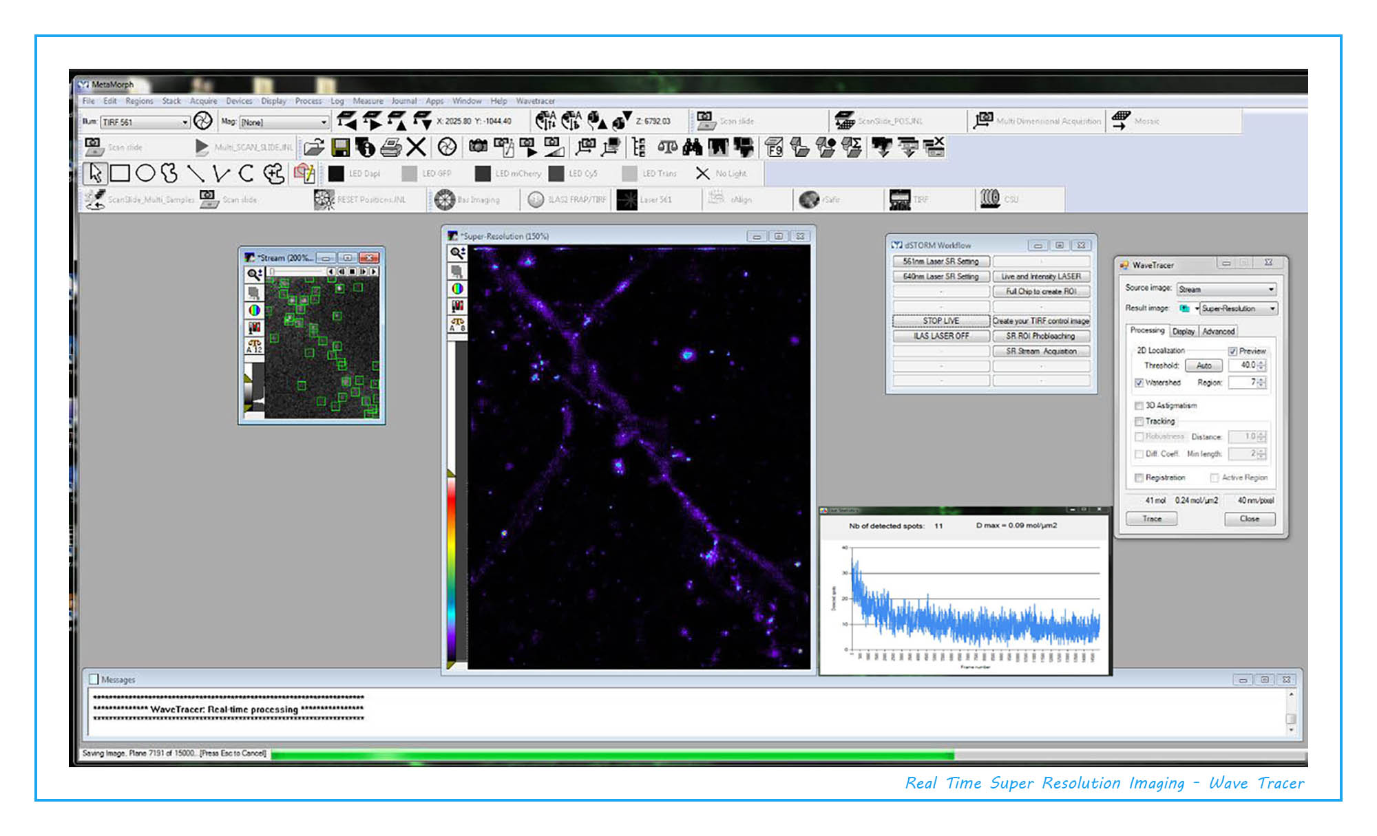
With synchronized image acquisition and processing, Wave Tracer MetaMorph software allow you to analyze details object on the nanometer scale, capturing organization of sub-cellular structures not readily discernible with conventional confocal or TIRF microscopy.
Super Resolution module taking full advantages of the powerful and flexible MetaMorph software capabilities. For added flexibility, a unique hardware acceleration component supports fast acquisition and analysis with real-time super-resolution image display. High resolution images can now be processed and displayed in real-time preventing researchers to wait to view their results until all images have been processed post acquisition. The system is compatible with most fluorescence and TIRF microscopes.
Super Resolution module taking full advantages of the powerful and flexible MetaMorph software capabilities. For added flexibility, a unique hardware acceleration component supports fast acquisition and analysis with real-time super-resolution image display. High resolution images can now be processed and displayed in real-time preventing researchers to wait to view their results until all images have been processed post acquisition. The system is compatible with most fluorescence and TIRF microscopes.
• User friendly interface
• Supports many different localization microscopy techniques
• Compatibility with many commercially available microscopes, cameras, laser launches, TIRF optics…
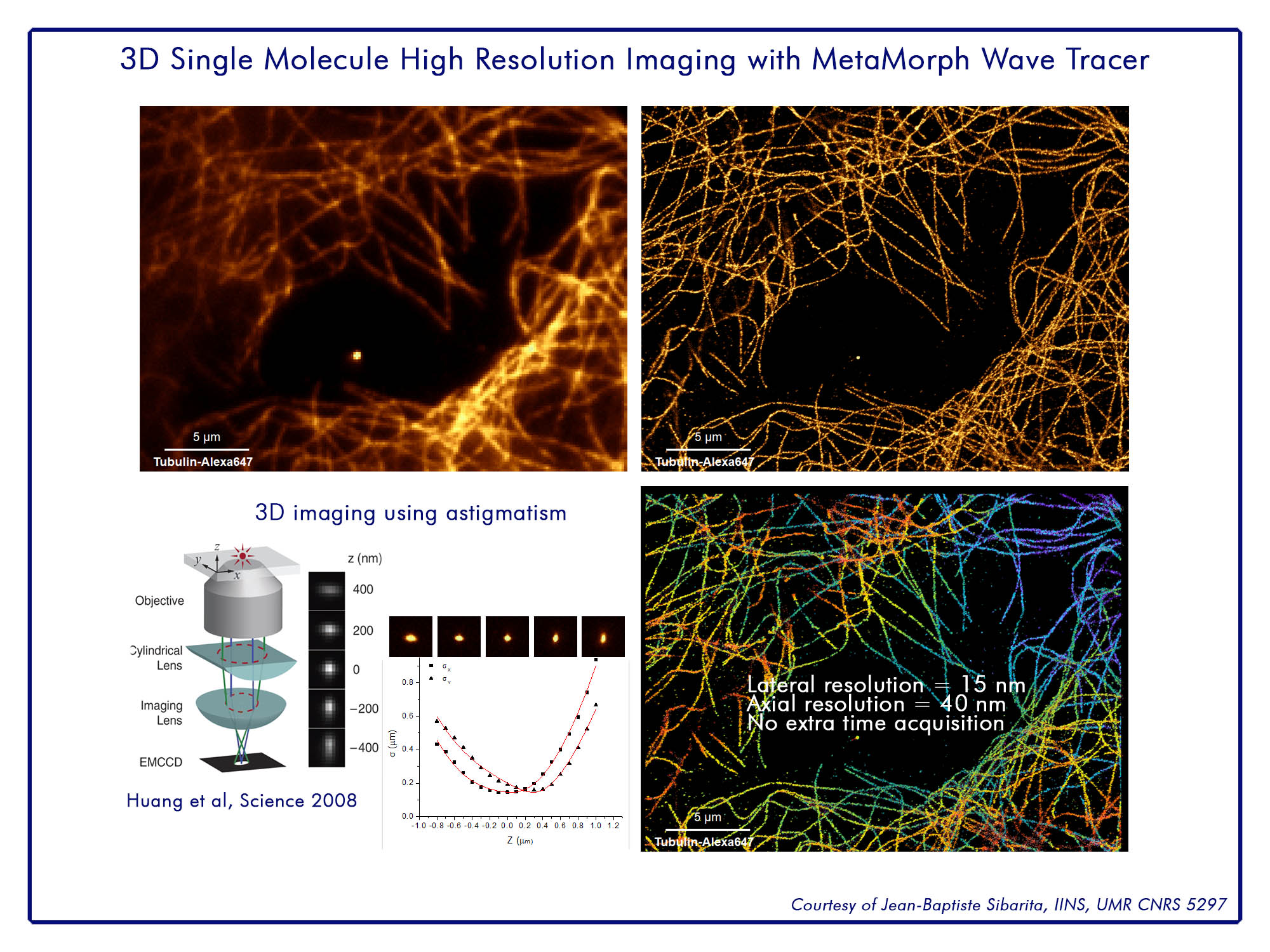
• 20 nm lateral / 40 nm axial with 3D astigmatic lens
• Streaming devices capabilities
• Real-time localizations, saving, & feedback
• Automatic laser feedback and adjustment
• Optimized workflow (when your image acquisition completes, the localization analysis and storage is also complete).
• Single molecule tracking
• Accelerated GPU processing
• 3D super resolution
• Supports many different localization microscopy techniques
• Compatibility with many commercially available microscopes, cameras, laser launches, TIRF optics…
• 20 nm lateral / 40 nm axial with 3D astigmatic lens
• Streaming devices capabilities
• Real-time localizations, saving, & feedback
• Automatic laser feedback and adjustment
• Optimized workflow (when your image acquisition completes, the localization analysis and storage is also complete).
• Single molecule tracking
• Accelerated GPU processing
• 3D super resolution
Could be enabled on any previously-installed hardware platforms (high-end video-microscope, spinning disk set up …)
Difficulties to do STORM, lack of laser power on your Super Resolution platform, absence of real-time analysis with Wave-Tracer, we have the solution, we can propose an upgrade of your set up.
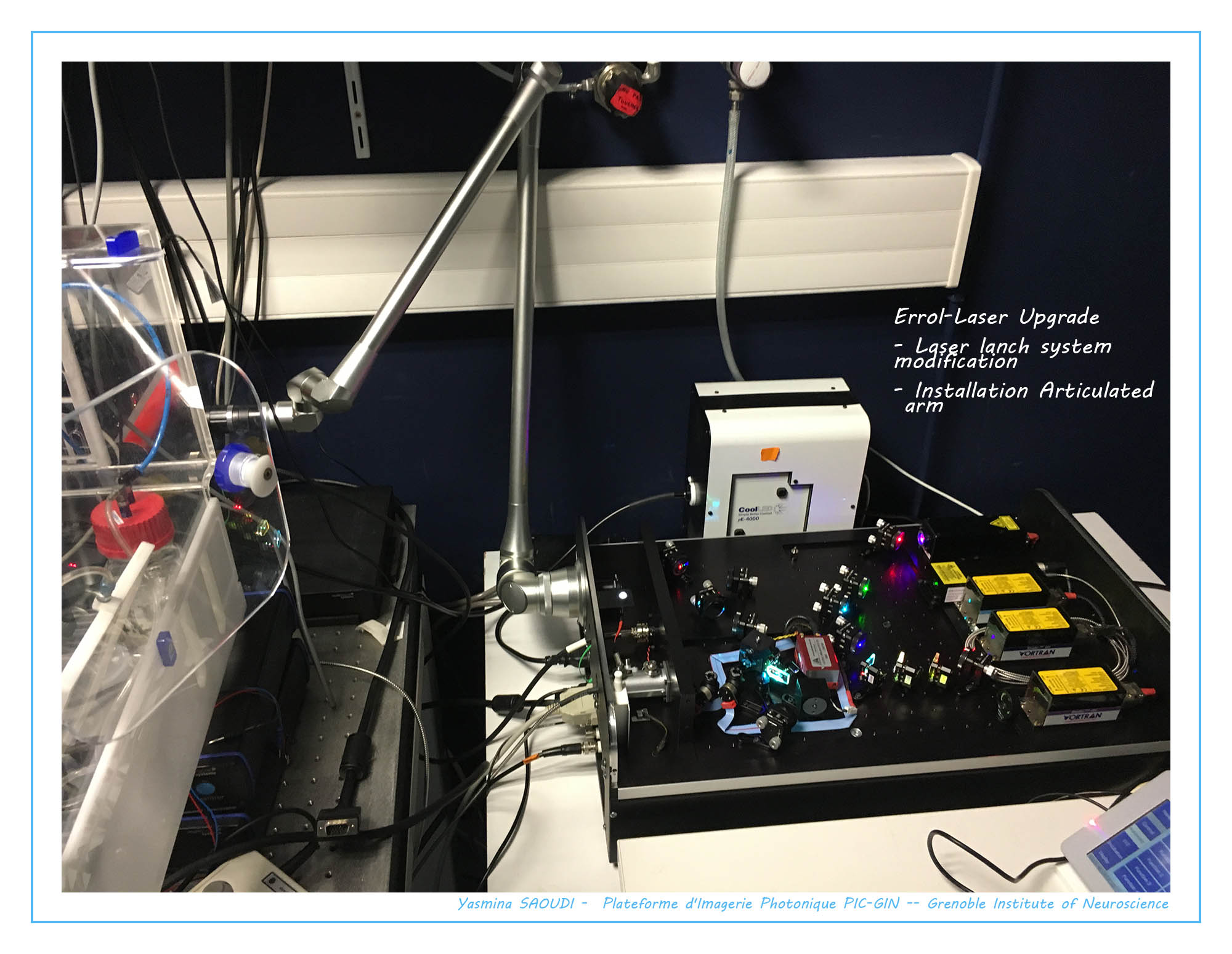
ZEISS CELL OBSERVER WITH SPINNING DISC CONFOCAL MICROSCOPE
Location
Grenoble Institut des Neurosciences, Inserm U1216 – Université Grenoble Alpes - Photonic Imaging Center (PIC GIN)
Bâtiment Edmond J. Safra, Chemin Fortuné Ferrini
38700 La Tronche
Contact person: Ms Yasmina SAOUDI
✉ yasmina.saoudi@univ-grenoble-alpes.fr
Applications
Fluorescence imaging, Confocal imaging, TIRF, dSTORM, PALM
Specifications
Inverted microscope capable of confocal imaging
• 100x NA: 1.46
• CSU-W1 Spinning disc
• 2 Flash 4.0 Hamamatsu cameras
• Lasers: 405nm – 488nm – 561nm – 640nm
• Errol-Laser: Laser launch modification (TIRF optical articulated arm)
• Metamorph – Wave Tracer v1.6 – Customized Super Resolution Taskbar