Systems / Confocal Microscopy Solution
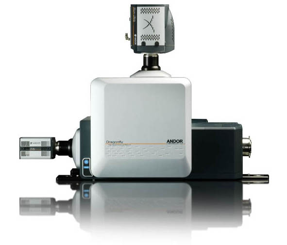
Dragonfly
Large Field of View Deeper
Getting the Most from Your Imaging

![]() Wide field of View
Wide field of View
![]() Deep tissue imaging
Deep tissue imaging
![]() Borealis perfect illumination delivery
Borealis perfect illumination delivery
![]() Powered by MetaMorph software platform
Powered by MetaMorph software platform
The Game-Changer In Confocal Microscopy
With The Andor Dragonfly, You Can Image At An Unrivalled Combination Of Speed, Sensitivity, Confocality And Resolution Beyond The Diffraction Limit
Larger field of view matching large sCMOS sensors | Capture more in a single image
Powered by MetaMorph software | Expand DragonFly multi-modal imaging capabilities & performance


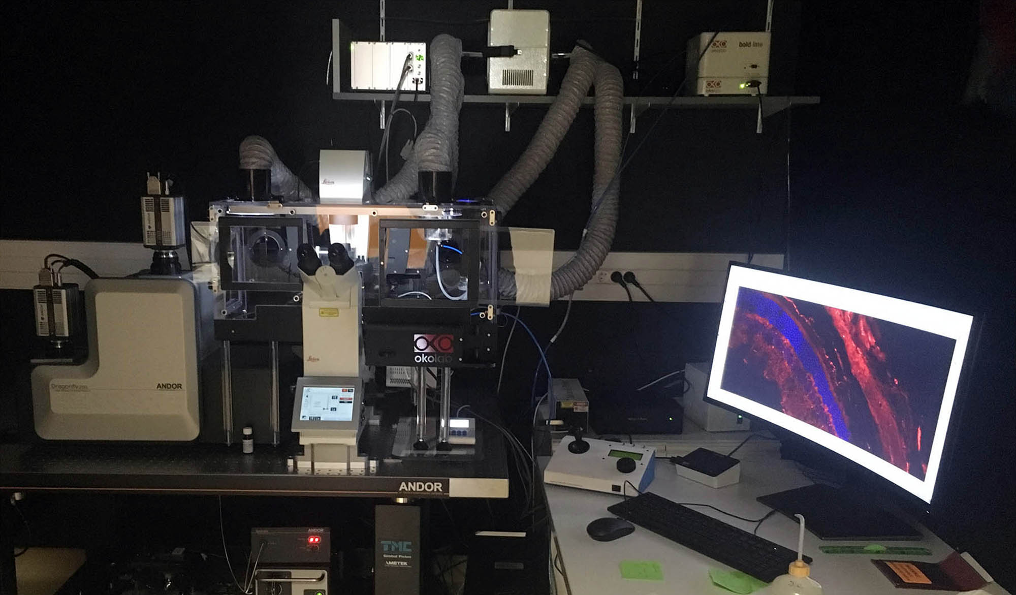
SPINNING DISC CONFOCAL MICROSCOPE – DRAGONFLY POWERED BY METAMORPH
Location
Grenoble Institute of Neuroscience
Eq « Central nervous system: From development to repair »
Bâtiment Edmond J. Safra – Chemin Fortuné Ferrini – 38700 La Tronche – FRANCE
Grenoble Institute of Neuroscience
Eq « Central nervous system: From development to repair »
Bâtiment Edmond J. Safra – Chemin Fortuné Ferrini – 38700 La Tronche – FRANCE
Contact person: Ms Homaira NAWABI ✉ homaira.nawabi@univ-grenoble-alpes.fr
Applications
Fluorescence imaging, Confocal imaging
Specifications
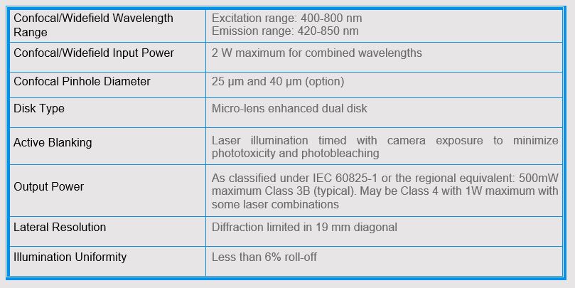
Inverted microscope capable of confocal imaging, equipped with immersion objectives.
![]() DragonFly 202 Spinning disc
DragonFly 202 Spinning disc
![]() Zyla 4.2 Andor camera
Zyla 4.2 Andor camera
![]() Andor ILE with 4 Lasers: 405nm (100mW) – 488nm (150mW) – 561nm (150mW) – 637nm (140mW)
Andor ILE with 4 Lasers: 405nm (100mW) – 488nm (150mW) – 561nm (150mW) – 637nm (140mW)
![]() Andor MicroPoint
Andor MicroPoint
