Systems / Confocal Microscopy Solution
CSU-X1 spinning Disk Confocal
Faster, Brighter, and More Versatile
GET IDEAL IMAGE QUALITY
SAVING CELLS FROM PHOTOTOXICITY or BLEACHING
![]() Faster
Faster
![]() Brighter
Brighter
![]() More versatile
More versatile
![]() Powered by MetaMorph software platform
Powered by MetaMorph software platform
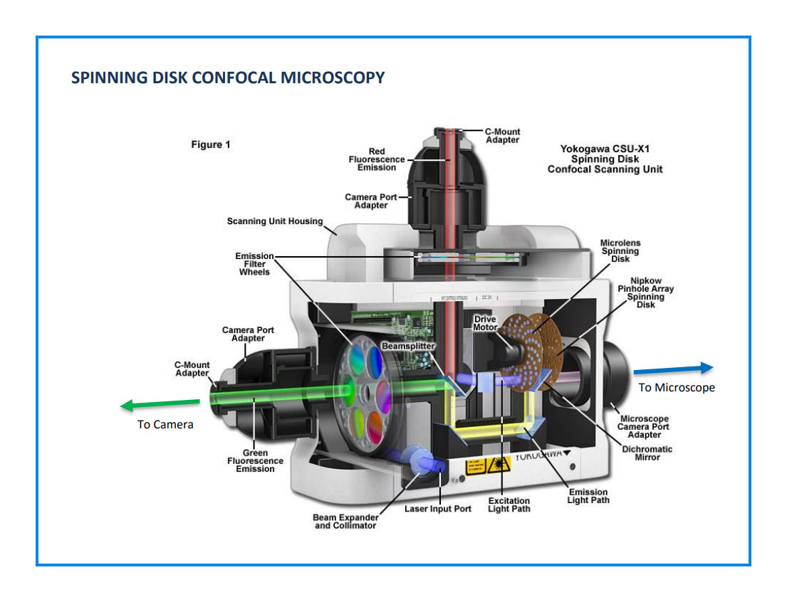
Schematic cut-away diagram showing a Yokogawa Electric Corporation spinning disk confocal unit.
This unit includes the motorized spinning pinhole disk, as well as a micro-lens disk and can be mounted to the camera port of a microscope to provide confocal images. The unit pictured can accommodate two cameras observing different wavelength channels, due to the internal dichroic beam splitter and filter wheels.
From Zeiss Campus: http://zeiss campus.magnet.fsu.edu/articles/spinningdisk/introduction.html
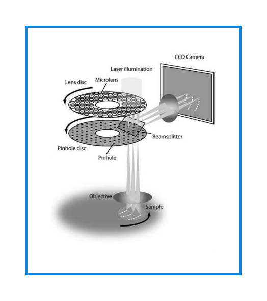
Microlens disk focuses illumination light through pinholes of primary disk. Returning light from the sample is separated by a dichroic mirror to scan across the sensor of a CMOS or CCD camera.
From Graf, R, J Rietdorf, and T Zimmermann. 2005. “Live Cell Spinning Disk Microscopy.” Adv Biochem Engin/Biotechnol 95: 57-75.
 #
#
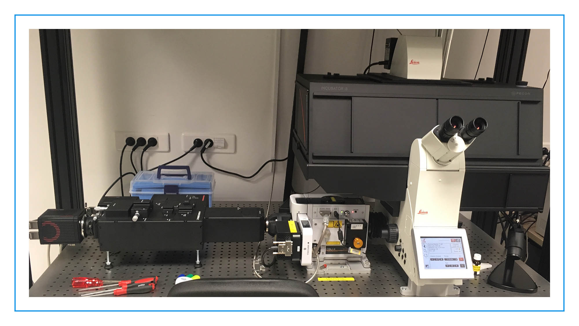
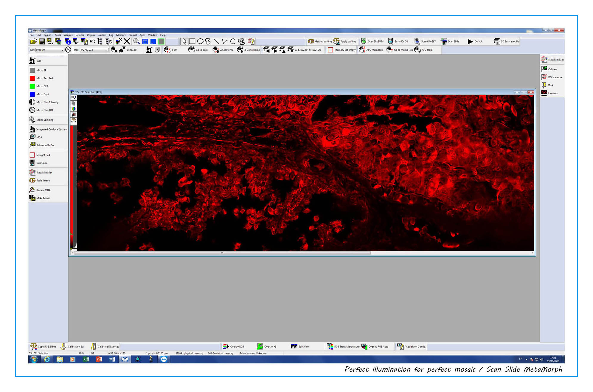
spinning disc confocal set up
NeurImag imaging facility – INSERM U894 – IPNP
102-108 rue de la Sante, 75014 Paris
Contact person: Ms Lydia DANGLOT ✉ lydia.danglot@inserm.fr
Applications
Fluorescence imaging, Confocal imaging
Specifications
Inverted microscope capable of confocal imaging, equipped with glycerol immersion objectives.
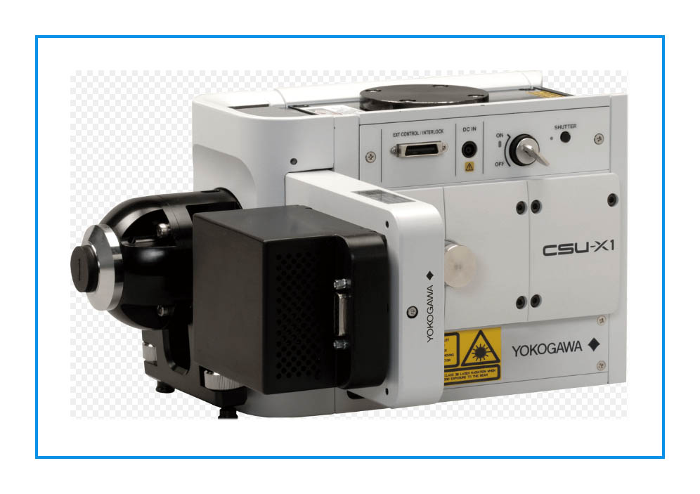
![]() CSU-X1 Spinning disc
CSU-X1 Spinning disc
![]() Hamamatsu Gemini-2C
Hamamatsu Gemini-2C
![]() 2 Flash 4.0 Hamamatsu cameras
2 Flash 4.0 Hamamatsu cameras
![]() Lasers: 405nm (200mW) – 488nm (180mW) – 561 (300 mW) – 640 (400 mW) – 730 (100 mW)
Lasers: 405nm (200mW) – 488nm (180mW) – 561 (300 mW) – 640 (400 mW) – 730 (100 mW)
![]() Errol-Laser STARSCAN
Errol-Laser STARSCAN